CenterEdgeAngleApp
The CE angle is one of the most frequently applied anatomic measures for the evaluation and description of the geometry of the hip in everyday orthopedic care. Measurements of CE angle in X-rays are valuable because you can determine and monitor objectively the severity of dysplasia, and establish the need for surgery and follow up the treatment.
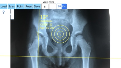
Wiberg’s or center edge (CE) angle is formed at the juncture of the Perkin line with line drawn from the center of the femoral head to the outer edge of the acetabular roof. The center edge angle may distinguish between acetabular insufficiency, under coverage or overcoverage of the femoral head by the acetabulum.
Measurements of CE angle in X-rays is important during the decision-making process for conservative or operative treatment, and follow up particular, in Developmental Dysplasia of the hip, or planning correction-osteotomies. Measuring angles in X-rays in clinical settings it is time consuming. Accessory instruments like protractors, goniometers, well sharped pencils, rulers must be available in a busy everyday practice. Usually you miss or you never had one or another. Also after measurement you have to compare the data that you measure with the normal reference values according to patient age and decide what could be considered normal in an X-ray of the hip and what is considered pathologic.
Center-Edge Angle app is a medical software aimed for orthopaedic surgeons, providing tools that allow doctors to:
-Securely import medical images directly from the camera or stored photos
-Offers a very convenient way to determine the most accurate possibly lines in order to measure the angles. By the aid of a circular transparent template, the points of interest are marked accurately. The automatically formed lines, drawn between points, measure automatically the angles of interest. The results are printed in degrees. By inputting the age, the measured angle is compared with values from normal reference database. In case the measured angle is beyond the normal range for that age, the hips are categorized as normal, borderline dysplastic, dysplastic or severe dysplastic or over coveraged, pincer type femuracetabular impingement (FAI) of the hip.
-Save the planned images, for later review or consultation.
All information received from the software output must be clinically reviewed regarding its plausibility before patient treatment! AI App is indicated for assisting healthcare professionals. Clinical judgment and experience are required to properly use the software. The software is not for primary image interpretation.
The app is a handy tool for an orthopaedic surgeon, radiologist, medical student or resident who wants objectively to monitor and determine the severity of dysplasia of the hip. The build-in comparison feature with the normal reference values according to patient age may help decide what could be considered normal or dysplastic or pincer (FAI). The app is not a simple goniometer, is an enhanced product which offers the ability to compare all the input data with medical reference database. The results are printed on the screen and the hips are categorized as normal or dysplastic or severe dysplastic or pincer (FAI) according to the angle measured. This feature it is particular useful especially in clinical settings where you need a quick results without losing time in looking for reference data according to age variations in huge textbook. The circular template to determine the points of interest and to mark them accurately are very useful in clinical settings where finding a sharpened pencil, a protractor and manage to draw with ruler lines over the patients x-rays is definitely a cumbersome and tedious task. You can load from your photo library or capture a photo from x-rays of the patient in you mobile phone or tablet, the App simply guides you to do the rest.


